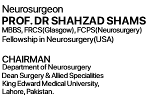
Prof. Shahzad Shams presently works as Head and Professor of Neurosurgery Department at King Edward Medical College/Mayo Hospital, Lahore, Pakistan.
Facts and Advice
Diseases Neurosurgeons Treat?
Neurosurgeons treat patients with tumors of the brain or spinal cord.
Accidents that lead to injuries of the head, spinal cord, or nerves require neurosurgical treatment.
Children born with a brain or spinal cord malformation at birth or with abnormal spinal fluid circulation require neurological surgery to help these children live a more normal life.
The most common condition that neurosurgeons treat is pain in the neck or lower back spreading to the arm or leg due to a ruptured disc. Slipped discs or pinched nerves may be treated non-surgically through bed rest, back braces, or physical therapy. Neurosurgery is performed to treat these patients when medicines fail to help or patients start to develop numbers or weakness of the affected limbs.
Neurosurgical Conditions
- Brain Tumours
- Brain Aneurysms
- Back Problems
- Neck Problems
- Brain Haemorrhage
- Endoscopic Neurosurgery
- Hydrocephalus
- Carpal Tunnel Syndrome
- Trigeminal Neuralgia
- Head Injuries
Brain Tumors
Brain Tumors Operated by Prof. Shahzad Shams:
Meningiomas, Malignant Brain Tumors(Glioma, Astrocytoma, Metastatic), Cerebellopontine angle tumors, Acoustic Neuroma Schwannoma Tumor, Pituitary tumor, Prolactinoma, Colloid cysts, Craniopharyngioma, Rathke’s Cleft Cyst, Sellar and Parasellar tumors and Brain Tuberculomas.
The surgery of various intracranial tumors is obviously one of the main areas of a neurosurgeon’s work. There is a large number of different tumours that can occur in the brain, arising from a variety of different tissues.
These include tumours of the nerve cells and their supporting structures, tumours of the lining membranes of the brain (meninges), various tumours associated with the pituitary gland, and tumours occurring on some of the cranial nerves (acoustic neuromas).
The brain can also be the site of tumour seedlings (metastases) from disease in other organs of the body.
Symptoms and Signs
Tumours can cause symptoms either by taking up space within the skull, leading to an increase in pressure within the head, or by interfering with the function of the adjacent brain or cranial nerves. Common symptoms therefore include headache, nausea and vomiting, epileptic fits or seizures or epilepsy and weakness or sensory loss in the face, arm or leg.
Imaging
Brain tumours can be imaged using CT or magnetic resonance imaging (MRI). Some types of tumour may then need surgical treatment, such as diagnostic biopsy or, where possible, complete surgical removal. Adjuvant treatments such as radio and chemo therapy may also be needed.
Results
The results of Brain tumor surgery are excellent specially after the Introduction of Endoscopes and endoscopy in brain surgery. Now it has become less invasive and hospital stay is less than 24 hours without any complications.
Brain Haemorrhage/Aneurysms/AVM’s
The commonest type of brain haemorrhage requiring neurosurgical treatment is a subarachnoid haemorrhage which is due to rupture of Brain aneurysms arising from Anterior communicating artery (Acom), middle cerebral artery (MCA), posterior communicating artery(Pcom), anterior cerebral artery(ACA), Internal Carotid artery(ICA) or AVM .
Haemorrhages (bleeding) within the brain can also be related to high blood pressure (hypertension) blood thinning (anticoagulant) medicines and some types of stroke.
Subarachnoid Haemorrhage
This type of haemorrhage usually occurs as the result of rupture of a small blister like abnormality (called an aneurysm) on one of the brain’s main arteries. Haemorrhage (bleeding) from one of these aneurysms is serious and potentially life threatening. It often presents with a very sudden and severe headache, which may be followed by loss of consciousness, nausea, vomiting or epileptic fits/seizures. The diagnosis is usually made with a CT scan or MRI Scan.
Once referred to a neurosurgical unit the blood vessels of the brain are imaged by Cerebral Angiography to see if there is an aneurysm or other abnormality present. If there is, this may need to be treated by a neurosurgical operation to put a Titanium metal clip across the neck of the aneurysm and thereby prevent further bleeding permanently from the ruptured aneurysm.
The commonest vessels involved are Anterior communicating artery, Middle cerebral artery , Posterior communicating artery and Internal Carotid Artery. The results of aneuyrsm surgery are excellent.
Results
The results of Brain Aneurysm clipping surgery are excellent.
Back Problems
Neurosurgeons treat a large number of problems with the lumbar spine (lower back). The more common ones include prolapsed intervertebral disc (slipped disc), spinal canal stenosis, and spinal tumours.
Prolapsed Intervertebral Disc (Slipped Disc)
The discs bulge backwards and press on a spinal nerve root. This can cause severe pain in the leg sciatica and backache, as well as weakness and sensory loss. Occasionally problems with the bladder and bowel may occur. The disc can be shown on magnetic imaging (MRI) and, if the symptoms are severe, can be removed by a microdiscectomy.
Spinal Stenosis
This means a progressive narrowing of the bony canal in the lumbar spine where the spinal nerves lie reffered as Lumbar Spondylosis or Lumbar Spondylitic changes. It is due to the gradual wearing out of the bones and ligaments as we get older. It can restrict the blood supply to the spinal nerves causing pain in the legs, as well as weakness and sensory changes, particularly after walking. The stenosis shows up on magnetic imaging (MRI) and can often be treated by an operation: lumbar laminectomy
Lumbar Spinal Tumours
Tumours can occur in the lumbar spine, although they are fortunately not common. They can cause low back and leg pain as well as leg weakness and sensory changes. Problems with the bladder and bowels can also occur. They are usually imaged with a magnetic resonance scan (MRI). Some may need to be removed surgically via a laminectomy.
Result
The results of Back surgery are excellent.
Neck Problems
Neurosurgeons treat a great many diseases of the cervical spine (neck). These include prolapsed intervertebral discs (slipped discs), canal stenosis (narrowing), and spinal tumours.
Prolapsed Intervertebral Disc (Slipped Disc)
The intervertebral discs bulge backwards in the neck and can press on the nerve roots going down the arm, causing severe arm pain, as well as weakness and sensory changes. A disc bulge can also press on the spinal cord itself, a potentially serious problem, which may cause weakness, sensory changes, alteration of bladder and bowel function, up to complete paralysis from the neck downwards. The disc is demonstrated using a magnetic resonance image (MRI). If necessary it can be surgically removed by an anterior cervical discectomy.
Cervical Canal Stenosis
The bones and ligaments of the cervical spine (neck) gradually wear out usually reffered as Cervical Spondylosis or Cervical Spondylitic changes . This causes the neck to lose some of its normal shape and the bony canal in which the spinal cord sits can become narrowed, sometimes quite severely.
This pressure on the spinal cord can be quite serious, causing weakness, sensory loss and bladder and bowel changes, or even complete paralysis below the neck. The stenosis can be demonstrated by magnetic resonance imaging (MRI) and, if necessary, can be surgically treated by a cervical laminectomy.
Cervical Spinal Tumours
Tumours can occur in the cervical spine (neck). These can cause pressure on the nerve roots supplying the arms, causing pain, weakness or sensory changes, as well as pressure on the spinal cord itself, causing weakness, sensory change, bladder and bowel disruption, or even complete paralysis. Tumours are visualised using magnetic resonance imaging (MRI) and some may need to be surgically removed, often via a cervical laminectomy.
Result
The results of Neck surgery are excellent.
Endoscopic Neurosurgery
I have now a clear belief that with the advent of Endoscopic neurosurgery and its advancement has greatly simplified the management of many intracranial ailments in adults and children. Similar in concept to other endoscopic surgery, intracranial neuroendoscopy reduces the surgical morbidity, shortens the hospital stay, and minimizes the cosmetic concerns associated with many major neurosurgical conditions.
In general, neuroendoscopy does not require large incisions on the scalp, removal of skull flaps, or extensive dissection through brain tissue. In the past several years the technological advancements in endoscope design have been substantial. A reduced size, improved resolution, and brighter illumination of the endoscope has allowed the benefits of endoscopic surgery to be applied in neurosurgery and it is definitely how the future neurosurgery would be performed.
Neuroendoscopy has dramatically altered the management of several diseases affecting the central nervous system of children and adults. At the centre of Minimal Access Neurosurgery at Omar Hospital, Jail road. Endoscopic neurosurgical procedures have been used in the treatment of pituitary tumours, hydrocephalus, intracranial cysts, intraventricular brain tumors, lumbar discectomy and decompression for lumbar spinal stenosis and Pediatric neurosurgery in children.
Result
The results of Endoscopic surgery are excellent.
Trigeminal Neuralgia
This is a condition, which may be treated by General Practioner’s(GPs), Neurologists, Neurophysicians, Psychiatrists and finally end up with Neurosurgeons. It is characterised by a severe spasmodic and lancinating pain, which affects one part of the side of the face. The pain can be excruciatingly severe and may be triggered by various actions, including chewing, swallowing, drinking hot or cold liquids, brushing the teeth, or being exposed to a cold wind.
The exact cause of the symptoms is not precisely known, but the pain usually affects one branch of the trigeminal nerve which supplies sensation to the side of the face. In many cases symptoms seem to be related to a loop of a blood vessel within the brain itself pressing on the trigeminal nerve. Such loops can sometimes be seen using magnetic resonance imaging (MRI).
Treatment
Endoscopic Microvascular Decompression – MVD
Endoscopic Microvasular decompression is a procedure in which a 2cm opening is made behind the ear and through this opening a endoscope of 4mm size is inserted to find the vessel which is offending the trigeminal nerve which is then separated from the nerve using micosurgical instruments and a small teflon graft is placed inbetween the vessel and nerve. Patient is immediately relieved of the severe pain on the face and is disharged within 24 hours from the hospital.
Result
The results for ENDOSCOPIC Microvascular decompression (MVD) is an excellent procedure and once done patient is relieved of the pain forever and permanently. Hospital stay is only for 24 hours.
Hydrocephalus
It means an accumulation of fluid within the brain, and a concomittent rise in pressure within the head. Some of the more common causes of hydrocephalus include aqueduct stenosis, normal pressure hydrocephalus, hydrocephalus secondary to haemorrhage or infection, benign intracranial hypertension and Arnold Chiari malformation.
Hydrocephalus can be investigated by a variety of means, including magnetic resonance imaging (MRI), to look at the ventricles within the head. Once the diagnosis has been confirmed, treatment may involve diverting the excess fluid from the brain to the abdomen by implanting a device called a VP SHUNT( Ventriculo-peritoneal shunt). These consist of a silicone tube, the flow along which is controlled by a valve.
There are many different varieties of these, some of which can have the valve’s pressure setting externally adjusted by the treating consultant using an electromagnet. Some types of hydrocephalus may be amenable to treatment with a neuro endoscope to create a drainage passage for the fluid within the brain itself.
More detail on hydrocephalus and ventriculo-peritoneal shunts can be found under the special topics menu.
Result
The results of VP Shunt surgery are excellent.
Carpal Tunnel Syndrome
Carpal tunnel syndrome is one of a number of entrapment neuropathies treated by neurosurgeons. This is a group of conditions, where symptoms occur when a nerve is “trapped” as a consequence of its normal anatomical path in the body becoming constricted. This compresses the nerve, causing tingling, weakness and often pain in the area supplied by that nerve.
The diagnosis can be confirmed by making electrical measurements of the speed of conduction along the nerve (nerve conduction studies). The neurosurgical treatment consists of a relatively small operation, often performed under local anaesthetic, whereby the fibrous carpal tunnel at the wrist is opened up in order to release the pressure on the nerve.
Result
The results of Carpal tunnel syndrome surgery are excellent.
Head Injuries
Neurosurgeons are involved in the management of all kinds of head injury, both minor and major. Surgical involvement is usually directed more towards the major head injuries.
Major Head Injuries
A major head injury can result in a variety of insults to the brain and skull. These include various types of skull fracture, large blood clots (haematomata) which may press on the underlying brain and damage it, contusions and lacerations to the brain itself, and diffuse bruising (swelling) of the entire brain.
These major injuries are often managed by emergency referral to a neurosurgical unit where life saving surgery may be needed to remove an intracranial blood clot (subdural or extradual haematoma), or to elevate and repair major skull fractures.
Patients with major head injuries may require time on intensive care and may also have their intracranial pressure monitored with a surgically implanted probe.
Minor Head Injuries
Neurosurgical involvement often consists of careful observation and/or CT scanning in order to exclude any of the more major injuries outlined above, and to be in a position to respond rapidly should any of these occur.
Result
The results of Minor and moderate head injury are excellent.


