Prof Shahzad Shams presently works as Chairman and Head Of Neurosurgery Department at King Edward Medical University & Mayo Hospital, Lahore.
Following Modern Techniques are used by him
- Endoscopic minimally invasive keyhole brain surgery
- Endoscopic minimally invasive keyhole removal of brain tumors through nose.
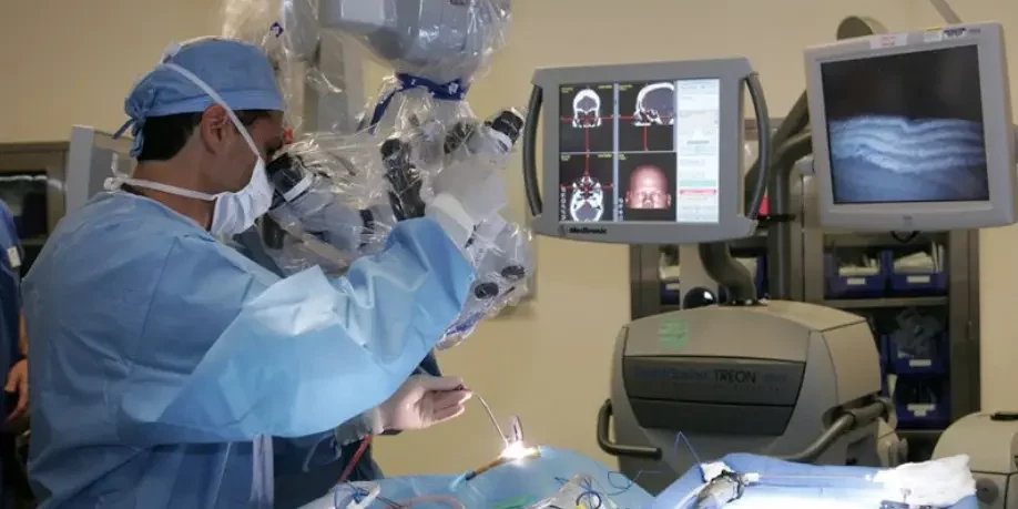
- Computer Assisted Neuronavigation Brain Tumor surgery used to precisely target the lesion and for complete removal.
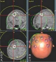
- Stereotactic Brain Tumor surgery a minimally invasive keyhole surgery technique for deep seated lesions with 1 mm accuracy
- High Powered Microscopic Brain Tumor surgery are used to magnify the tumor for safe removal .
- Endoscopic surgery ETV for Hydrocephalus attached to camera with images on monitor.
- Per operative neurophysiological monitoring is done to protect important nerves and areas of Brain.
- Safest and best technique performed by only one Team of Prof Shahzad Shams in Pakistan called Translabyrinthine minimally invasive keyhole surgery and approach for Acoustic Neuroma surgery.
All these methods help in safe and complete removal of tumors giving excellent results.
About Stereotactic Brain Biopsy done through Keyhole surgery
Prof Shahzad Shams is the first Neurosurgeon in Punjab and KPK to start Stereotactic neurosurgery in the Private sector which involves mapping the brain in a three-dimensional coordinate system.
With the help of MRI and CT scans and 3D computer workstations, he is able to accurately target any area of the brain in stereotactic space (3D coordinate system). He uses RM frame system and Incomed IPS Planning software for this purpose.
Stereotactic brain biopsy is a minimally invasive procedure that uses this most advanced and cutting edge technology to obtain samples of brain tissue for diagnostic purposes and for aspiration of cystic lesions.
Indications
This procedure is used to obtain tissue samples of areas within the brain that are suspicious for tumors or infections. The main indications for stereotactic biopsy are :-
- Deep-seated lesions
- Lesions in Eloquent areas of brain like Thalamus and Brain Stem.
- Multiple lesions like Metastatic lesions
- Diffuse Infiltrative lesions like Encephalitis and Diffuse Glioma
- Lesions in patients who cannot tolerate anesthesia like old aged
- For Aspiration of Brain abscess and cystic lesions
- For Aspiration of hematomas

Technique
On the afternoon of surgery a head ring is placed on the patient. This involves giving local anesthesia to the skin in four areas and placing the ring on the head with four pins. A CT scan is then performed.
In the operating room, the patient receives light sedation. An incision only a few millimeters long is made in the scalp and a small hole is drilled into the skull. A thin biopsy needle is inserted into the brain using the coordinates obtained by the computer workstation.
The specimen is then sent to the pathologist for evaluation. Patients are monitored for several hours following the procedure and usually go home the same day.
Accuracy of the RM frame and IPS Planning system is of sub millimeter level and is considered to be the best system in the world giving 100% results to reach diagnosis.


